 rganauta
Inside the retina in 3D
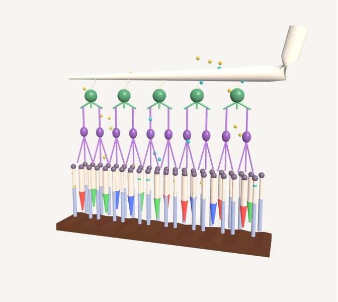
The retina has layers of neurons stacked on top of each other. Light comes in from the top, crosses the ganglion cells (green) and the bipolar cells…
Explained for every age
For kids (7–11)
Here you see the retina, the layer at the back of the eye, as if you were looking at it through a giant microscope. The little yellow balls are light coming down from the top: it goes through several layers of cells and reaches the bottom, where the cones and rods are. They notice the light and send a signal, the little light-blue glow, that travels up to the white cables that go to the brain.
For teens (12–16)
The retina has layers of neurons stacked on top of each other. Light comes in from the top, crosses the ganglion cells (green) and the bipolar cells (purple), and reaches the photoreceptors: rods and cones. They turn light into an electrical signal, which travels back up: it passes to the bipolar cells and then to the ganglion cells, whose axons form the optic nerve. Each eye has about 100 million rods and about 6 million cones. Underneath everything is the pigment epithelium, a dark layer that takes care of the photoreceptors.
Pre-Med (17+)
The vertebrate retina is inverted: light passes through the ganglion cell layer and the inner nuclear layer before reaching the outer segments of the photoreceptors, which rest on the pigment epithelium. Depending on the source, each retina has roughly 92 to 120 million rods and about 4.6 to 7 million cones, but only 0.7 to 1.5 million ganglion cells, so on average each ganglion cell receives information from about 100 photoreceptors. Photoreceptors, bipolar cells and horizontal cells work with graded potentials, as do most amacrine cells; the action potentials that travel along the optic nerve are generated by the ganglion cells (and, within the retina, only a few amacrine cells also produce them). The scene is a diagram, not to scale, and omits horizontal and amacrine cells.
What you can explore
Rod cellCone cellRetinal bipolar cellRetinal ganglion cellRetinal pigment epitheliumVisual phototransduction
Missions
- Light's journey Explorer
- Layers of the retina Discoverer
- Photoreceptors and the clinic Pre-Med