 rganauta
Liver lobule in 3D
The liver lobule is the microscopic unit of the liver: a roughly hexagonal prism with a central vein in the middle and a portal triad at each corner.…
Explained for every age
For kids (7–11)
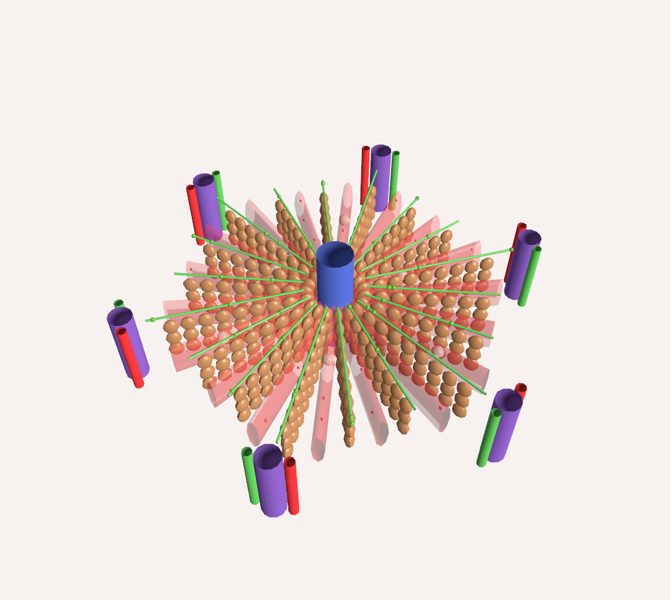
The liver is made of thousands and thousands of tiny pieces shaped like hexagons, like the cells of a honeycomb. Here you see one of them, magnified a lot. Blood comes in at the corners, passes between rows of liver cells that clean it, and leaves through the blue tube in the center. At the same time, those cells make bile, a green juice that travels the opposite way, toward the corners.
For teens (12–16)
The liver lobule is the microscopic unit of the liver: a roughly hexagonal prism with a central vein in the middle and a portal triad at each corner. Plates of hepatocytes run from the center to the edges, and between them run the sinusoids, wide capillaries where blood from the portal vein (about 75% of the flow, rich in nutrients absorbed in the intestine) mixes with blood from the hepatic artery (the remaining 25%, rich in oxygen). Blood moves toward the central vein; bile, on the other hand, travels through canaliculi toward the bile ductules at the corners. In the scene, the red and purple particles are blood and the green ones are bile.
Pre-Med (17+)
Classic lobule: a hexagonal prism centered on the central vein (terminal hepatic venule), with portal tracts at the vertices that contain a branch of the portal vein, a hepatic arteriole, one or two bile ductules lined by cuboidal epithelium, lymphatics and nerve branches. The portal vein supplies about 75% of the flow and the hepatic artery about 25%, but each covers roughly half of the oxygen demand. Mixed blood runs through the sinusoids from the periphery to the center, drains into the central vein and, via the hepatic veins, into the inferior vena cava. Bile flows the opposite way: from the canaliculi to the canals of Hering and the portal ductules. For function and pathology, the hepatic acinus is used, centered on the axis between two portal tracts and divided into three zones. The scene is a diagram, not to scale.
What you can explore
Liver sinusoidCentral veinPortal triadBile canaliculusKupffer cell
Missions
- The liver's filter Explorer
- Blood in, bile out Discoverer
- Acinus, sinusoids and liver injury Pre-Med